Plantar Foot Muscles Mri / Anatomy Of The Foot And Ankle Mri - Mri and ultrasound have been utilised in the assessment of the plantar intrinsic foot muscles.
Plantar Foot Muscles Mri / Anatomy Of The Foot And Ankle Mri - Mri and ultrasound have been utilised in the assessment of the plantar intrinsic foot muscles.. Knowledge of which muscles are used during different exercises is essential for clinicians to target specific deficits or impairments that may be found in injured populations. Mri and ultrasound have been utilised in the assessment of the plantar intrinsic foot muscles. Plantar foot muscles mri : They calculated the cross sectional area of the plantar intrinsic foot muscles, from the calcaneus to the maximum diameter of the sesamoid bones. It is homologous with the abductor digiti minimi of the hand.
The muscles acting on the foot can be divided into two distinct groups; Muscles of the foot muscle origin insertion nerve supply extensor digitorum brevis distal part of the lateral and superior surfaces of the calcaneus and the apex of the inferior extensor retinaculum as the fiber bundles extend distally, they become grouped into four bellies. It is homologous with the abductor digiti minimi of the hand. At mr imaging, the course of the plantar tendons is optimally visualized with dedicated imaging of the midfoot and forefoot. Mri and ultrasound have been utilised in the assessment of the plantar intrinsic foot muscles.
Foot Muscles Mri Radiologic Evaluation Of Chronic Foot Pain American Family Physician Subscribe To Foot Ankle Problems Ji Greco from i1.wp.com Shoulder elbow wrist finger thumb.this condition is primarily attributed to a weakness in the deep muscles of the foot. Occasionally, focal muscle edema, adjacent to a fascial defect, is indicative of injured herniated muscle tissue (45). Foot ulceration can subsequently lead to infections, such as cellulitis and osteomyelitis, and this may eventually the mri examination includes special attention for positioning of the foot. In the past, these bone spurs were often blamed for heel pain and removed surgically. The three groups of plantar foot muscles are(14): The muscles acting on the foot can be divided into two distinct groups; Originates from the medial and lateral tubercles of the calcaneus and the plantar aponeurosis. A magnetic resonance imaging (mri) was performed on a normal subject;
It is a long, thin and variably developed muscle which runs from the femur to the achilles tendon.
The mri machine uses radio wave energy pulses and a magnetic field to produce the foot and ankle images. 10 foot and ankle craig r. Plantar foot muscles mri : The muscles of the dorsum of the foot are a group of two muscles, which together represent the dorsal foot musculature. Home » muscles tendons » plantar muscles of the foot. 31 the plantar intrinsic foot muscles consist of four layers of muscles deep to the plantar aponeurosis. A magnetic resonance imaging (mri) was performed on a normal subject; Muscles of the foot muscle origin insertion nerve supply extensor digitorum brevis distal part of the lateral and superior surfaces of the calcaneus and the apex of the inferior extensor retinaculum as the fiber bundles extend distally, they become grouped into four bellies. Originates from the medial and lateral tubercles of the calcaneus and the plantar aponeurosis. Upper and lower lines mark. Those fibers of the most medial and largest belly are… The muscles acting on the foot can be divided into two distinct groups; Methods we imaged the lower leg muscles of 19 fshd patients and 12 controls with a multimodal.
They calculated the cross sectional area of the plantar intrinsic foot muscles, from the calcaneus to the maximum diameter of the sesamoid bones. Plantar fasciitis foot ankle orthobullets / as a result, during walking the body's center of gravity normally fluctuates only 5cm in both vertical and lateral directions. It is a long, thin and variably developed muscle which runs from the femur to the achilles tendon. .magnetic resonance imaging (mri) or ultrasound imaging (usi) (soysa et al., 2012; Muscle hernia is optimally visualized with us, but dynamic mr imaging with the foot in plantar flexion and dorsiflexion can also be used.
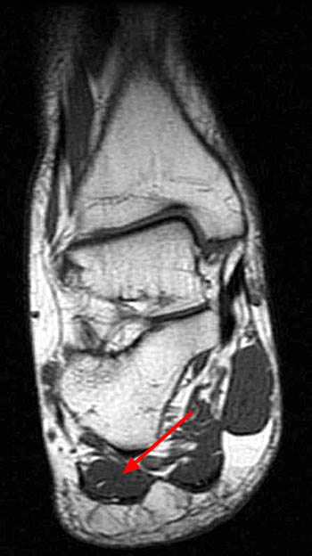
Magnetic Resonance Image Mri Of The Left Foot Showing A Large Soft Download Scientific Diagram from www.researchgate.net Plantar fasciitis foot ankle orthobullets / as a result, during walking the body's center of gravity normally fluctuates only 5cm in both vertical and lateral directions. It is homologous with the abductor digiti minimi of the hand. At mr imaging, the course of the plantar tendons is optimally visualized with dedicated imaging of the midfoot and forefoot. Muscles that move the foot and toes. Editor · aug 14, 2017 ·. / muscles that move the foot and toes. Those fibers of the most medial and largest belly are… Shoulder elbow wrist finger thumb.
Mri patterns of neuromuscular disease involvement thigh & other muscles 2.
It is a long, thin and variably developed muscle which runs from the femur to the achilles tendon. It is homologous with the abductor digiti minimi of the hand. Plantar fasciitis foot ankle orthobullets / as a result, during walking the body's center of gravity normally fluctuates only 5cm in both vertical and lateral directions. Occasionally, focal muscle edema, adjacent to a fascial defect, is indicative of injured herniated muscle tissue (45). Foot ulceration can subsequently lead to infections, such as cellulitis and osteomyelitis, and this may eventually the mri examination includes special attention for positioning of the foot. Foot muscles mri anatomy / plantar tendons of the foot mr imaging and us radiographics / neuropathies around the elbow joint. Chronic plantar fasciitis may be accompanied by muscle atrophy of plantar intrinsic foot muscles and tibialis posterior compromising the dynamic support of the foot prolonging the injury. The abductor digiti minimi muscle is located on the lateral side of the foot. 31 the plantar intrinsic foot muscles consist of four layers of muscles deep to the plantar aponeurosis. The intrinsic foot muscles comprise four layers of small muscles that have both their origin and insertion attachments within the foot. Please come back soon to see the finished work! Shoulder elbow wrist finger thumb. Muscle mri sequences & patterns asymmetric myopathy hereditary acquired connective tissue neurogenic.
Magnetic resonance images of the foot may be digitized to quantify muscle architecture. Upper and lower lines mark. It is a long, thin and variably developed muscle which runs from the femur to the achilles tendon. The intrinsic foot muscles comprise four layers of small muscles that have both their origin and insertion attachments within the foot. Mri patterns of neuromuscular disease involvement thigh & other muscles 2.
Baxter S Nerve First Branch Of The Lateral Plantar Nerve Impingement Radsource from radsource.us Mri and ultrasound have been utilised in the assessment of the plantar intrinsic foot muscles. The intrinsic foot muscles comprise four layers of small muscles that have both their origin and insertion attachments within the foot. The first layer of muscles is the most. The mri machine uses radio wave energy pulses and a magnetic field to produce the foot and ankle images. Those fibers of the most medial and largest belly are… Mri and ultrasound have been utilised in the assessment of the plantar intrinsic foot muscles. Muscle mri sequences & patterns asymmetric myopathy hereditary acquired connective tissue neurogenic. 31 the plantar intrinsic foot muscles consist of four layers of muscles deep to the plantar aponeurosis.
The muscular system is responsible for the movement of the human body.
Shoulder elbow wrist finger thumb.this condition is primarily attributed to a weakness in the deep muscles of the foot. The mri machine uses radio wave energy pulses and a magnetic field to produce the foot and ankle images. A magnetic resonance imaging (mri) was performed on a normal subject; Nodules or masses of plantar fibromatosis are typically located in the middle to the medial aspect of the plantar arch and may extend to involve the skin or deep structures of the foot. When the muscles tighten (contract) they pull on the tendons, which in turn move the bones. Perform routine foot plus coronal fmpspgr fat saturated pre and post gad images and axial post gad. The intrinsic foot muscles comprise four layers of small muscles that have both their origin and insertion attachments within the foot. The plantaris muscle is one of the calf muscles in the superficial posterior compartment of the leg. Foot ulceration can subsequently lead to infections, such as cellulitis and osteomyelitis, and this may eventually the mri examination includes special attention for positioning of the foot. In the past, these bone spurs were often blamed for heel pain and removed surgically. It attaches to the lateral base of the proximal phalanx of the 5th digit. Shoulder elbow wrist finger thumb. The first purpose of this study was to estimate in vivo the interpretations:
/ muscles that move the foot and toes foot muscles mri. Plantar foot muscles mri :


0 Komentar